Previous work

Fabric cancer cell by Kath Howard (2013)

Previous work on display by Kath Howard (2013)

Previous work on display by Kath Howard (2013)
When looking back at my work from the beginning of the year, I concluded that I needed to look into a way of making my fabric disease cells look more aesthetically pleasing, which would in turn make viewers of my artwork want to hold them. When making my previous pieces, the materials I used prevented me from doing this, causing my pieces to look messy and unattractive. At the beginning of the year I had planned to create a large collection of fabric disease cells, however I feel that with the time I’ve been given to create these pieces I would rather make only a few pieces in which I would spend more time on in order to make them looked detailed and aesthetically pleasing. Although I was unhappy with the over all turnout of my previous work, I was still quite fond of one fabric disease cell in particular which I had spent more time on and for this reason, I decided to keep this particular piece to display with my upcoming work.
Materials

Velvet materials in various colours. Photo by Kath Howard (2014)

Assortment of coloured cotton material. Photo by Kath Howard (2013)

Polyester stuffing. Photo by Kath Howard (2014)

Assortment of blue sequins. Photo by Kath Howard (2014)

Assortment of 4mm pearl beads for decoration. Photo by Kath Howard (2014)

Coloured cotton thread. Photo by Kath Howard (2014)
For me, changing my choice of materials was an important part of improving my work. From working mainly with cotton materials, I changed to using velvets as much as possible in order to make my pieces soft and pleasant to touch. However, I also kept to using some cotton material in order to create different textures and qualities in each piece. When choosing my fabric, I kept with the idea of using a range bright, non-offensive colours as much as possible in order to make my pieces interesting. As well as fabric, I also chose to use sequins and beads to add more detail to my pieces. I also felt like using these types of materials would draw more attention to them as well as adding more textures and colour.
Drawings

‘Crookes hyaline a x1000’ by Kath Howard (2013) image used for sketches.

Sketch of disease cells by Kath Howard (2014)

‘HaemangioblastomaHP’ image by Kath Howard (2013) used for sketches.

Watercolour sketch by Kath Howard (2014)

Breast cancer cell sourced from web and used for sketches.

Sketch of breast cancer cell by Kath Howard (2013)
Before beginning to make my pieces, I created a few rough sketches with inspiration from my own images of disease under the microscope as well as found images from the Wellcome Science Photo Library. When drawing out sketches, I though about shape, colours and textures as well as possible materials. Looking back at my previous project, I chose a sketch I had made at the beginning of the year and decided to finally create a piece based on this sketch. I found that drawing out my ideas helped, as I find that when working with materials, the outcome is never as accurate as the image being replicated. However, I still found that my finished pieces changed whilst in the process of making.
Making

Making the fabric disease cells. Photo by Kath Howard (2014)

Making the fabric disease cells. Photo by Kath Howard (2014)

Making the fabric disease cells. Photo by Kath Howard (2014)

Making the fabric disease cells. Photo by Kath Howard (2014)

Making the fabric disease cells. Photo by Kath Howard (2014)

Making the fabric disease cells. Photo by Kath Howard (2014)

Making the fabric disease cells. Photo by Kath Howard (2014)

Making the fabric disease cells. Photo by Kath Howard (2014)

Making the fabric disease cells. Photo by Kath Howard (2014)
To begin making my final pieces, I created the main body of the disease cells using white and light pink velvet. I created several different size bodies in which to work with and add detail to and then stuffed them with polyester white stuffing to make the pieces soft to touch and squeeze. When making the fabric disease cells, I followed to my sketches and images as much as possible, cutting out my desired shapes of fabric and sewing neatly onto the body. With one of my pieces, I created different sized stuffed balls of velvet in which I attached to the body of the cell. I felt this particular method added a desire to hold the fabric disease cell and play with the shape of the piece.

Making the fabric disease cells. Photo by Kath Howard (2014)

Making the fabric disease cells. Photo by Kath Howard (2014)

Making the fabric disease cells. Photo by Kath Howard (2014)

Making the fabric disease cells. Photo by Kath Howard (2014)

Making the fabric disease cells. Photo by Kath Howard (2014)

Making the fabric disease cells. Photo by Kath Howard (2014)
Once the main parts of the fabric disease cells were added, I began to add more delicate details such as sequins and beading to three of my four pieces. I decided against using beads and sequins on my older piece as I felt it was unnecessary due to it already being quite heavily textured and therefore I didn’t want to over do it. When securing the beads and sequins I made sure to keep them as neat as possible, and only selected particular areas of the pieces to place them in order to not make the fabric disease cells look over crowded or too kitsch.
Comparing fabric disease cells. Photo by Kath Howard (2014)
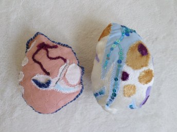
Making my final pieces turned out to be quite a long process in order to make them look as aesthetically pleasing as possible as well as making them resemble the images in which inspired them in the first place. However I was happy with my outcome and felt that my new fabric disease cells were a lot better made and represented my concept of how disease under the microscope is deceivingly beautiful much more.

One of the finished fabric disease cells by Kath Howard (2014)

Three of the finished fabric disease cells by Kath Howard (2014)
Video
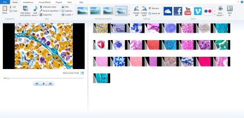
Screenshot of making the video. Photo by Kath Howard (2014)
To accompany my fabric disease cells, I created a loping video which consisted of my own images of disease under the microscope which I had sourced from visiting Derriford Hospital last year. When selecting the images, I chose the most interesting as well as aesthetically pleasing, selecting bright colours and complex patterns. When creating this video, my initial intentions were to include some unpleasant images of surgery relating to disease, or the results of cancer cells amongst the more beautiful images of diseases under the microscope, however, I feel that only showing the more pleasant images allows the viewer to interpret my fabric disease cells how they wish before knowing what they really are a representation of. I feel the video allows my viewers to make a connection between the 3D pieces and the images within the video, however I feel that it doesn’t give too much away, complimenting my fabric pieces well. I also feel that although these images are primarily for scientific purposes, they can be seen as artworks themselves, supporting my ideas about the collaboration between art and science within my work.
(Please use this link to visit the final video and artworks on display)
References: Wellcome, (n.d.). Breast cancer cell. [image] Available at: http://nutritionfacts.org/wp-content/uploads/2012/04/stem-cell-blog-for-NF-on-april-19th.jpg?c3d2dd [Accessed 1 Jun. 2014].


